- OTHM Level 4 Unit Managing Digital Information (J/650/3386) Assignment Brief 2026
- OTHM Level 4 Unit Computer and Network Technology (L/617/2268) Assignment Brief 2026
- OTHM Level 4 Unit Web and Mobile Applications (H/650/3385) Assignment Brief 2026
- OTHM Level 4 Unit Systems Analysis and Design (F/617/2266) Assignment Brief 2026
- OTHM Level 4 Unit Principles of Computer Programming (F/650/3384) Assignment Brief
- OTHM Level 4 Unit Cyber Security (D/650/3383) Assignment Brief 2026
- BSC072 Strategic Information Management Coursework Brief 2026 | Loughborough University
- Financial Econometrics Assessed Coursework 1 2026 | University of Portsmouth
- UHRINN304 Medicines Management Summative Assessment 2026 | University of Suffolk
- CIPD Level 7 Unit 7OS06 Wellbeing at Work Assignment Example 2026
- CIPD Level 7 Unit 7OS05 Managing People in an International Context Assignment Example 2026
- CIPD Level 7 Unit 7OS04 Advanced Equality, Diversity and Inclusion Assignment Example 2026
- 7FNCE037W Global Entrepreneurial Finance Assessment Briefing 2026 | UOW
- CIPD Level 7 Unit 7OS03 Technology Enhanced Learning Assignment Example 2026
- BSC006 Financial Trading Assessment Coursework Brief 2026 | Loughborough University
- CIPD Level 7 Unit 7OS01 Advanced Employment Law in Practice Assignment Example 2026
- CIPD Level 7 Unit 7LD03 Designing Learning to Improve Performance Assignment Example 2026
- CIPD Level 7 Unit 7LD02 Leadership and Management Development in Context Assignment Example 2026
- BUS6012 Governance, Law and Ethics Assessment Brief 2026 | Arden University
- CIPD Level 7 Unit 7LD01 Organisational Design and Development Assignment Example 2026
FHEQ level 5 BB2804: The coursework for this assessment block involves producing an A1 format, portrait orientation poster to present at the Brunel Symposium: Data Analysis, Interpretation and Presentation, Assignment, BUL, UK
| University | Brunel University London (BUL) |
| Subject | Interpretation and Presentation |
Description of assessment task
The coursework for this assessment block involves producing an A1 format, portrait orientation poster to present at the Brunel Symposium on Metabolism and Tumour Biology on the subject of the case study.
Your task will be to analyse, describe and interpret the clinical findings given in the case study using appropriate techniques. The introduction should briefly state what the poster is about in approximately 250 words. State what the subject is, why it’s important and give some background information. Use references to support your document. Use BLAST to analyse the sequence in figure 2 and identify the mutation in the sequence. The numerical data should be analysed and presented graphically along with any statistical analysis of the data you perform.
The photomicrographs should be labelled. The poster should contain 6 figures presenting the data with clear and concise figure legends you write yourself expressing your understanding of what is being shown and its meaning. The poster should conclude with a section on the observations made, the likely prognosis and treatments possible for the individual and conclusions that can be drawn. Any references used should be listed at the end of the poster. You will be assessed on the presentation of the poster, organization, clarity, as well as the analysis of the data. A 5-minute presentation and questions asked by a pair of examiners will also contribute to the overall mark.
Do You Need Assignment of This Question
Case study
In April 2019, a 48-year-old man was admitted to the department of Hepatobiliary Surgery at the local hospital due to upper abdominal discomfort and yellow-stained skin mucosa. Prior to hospital admission, the patient visited the local GP in three separate occasions (05/2018; 08/2018; 01/2019) over the past year manifesting a series of symptoms including mild yellow staining of the skin mucosa, a deep urine colour and nausea. During those visits he was given treatment for heartburn and gastroesophageal acid reflux with no obvious improvement. Blood samples were taken and analysed along with those taken over the past year in the three distinct occasions. The levels of two liver enzymes (ALT and AST) are given in Figure 1.
A contrast-enhanced computed tomography (CECT) scan was ordered. The results revealed a 14-mm nodule observed in the caudate lobe of the liver. The hepatic nodule was suspected of being a primary intrahepatic cholangiocarcinoma (ICC). To confirm this hypothesis a liver biopsy was performed.
Previous studies have shown that mutations of isocitrate dehydrogenase (IDH2) – a metabolic enzyme that converts isocitrate to alpha-ketoglutarate (α-KG) while reducing NADP+ to NADPH and liberating CO2 – occur in ICC. DNA was extracted from the liver biopsy of the ICC patient. Exon 4 of the IDH2 gene was amplified by PCR and sequenced. The sequence is shown in Figure 2.
Human cholangiocytes containing normal levels of unmutated IDH2 (endogenous protein; parental) were transfected with expression vectors either containing wild-type IDH2 cDNA or the mutated IDH2 cDNA extracted from the patient. The cells were grown in culture and IDH2 activity was measured two days after transfections after lysis of the cells. The activity of IDH2 was analysed through the reduction of NADP+ to NADPH, which was measured at 340 nm. Results of three independent measurements are shown in Figure 3.
Activation of the NF-kB transcription factors has been closely linked to the pathogenesis of ICC. Therefore, NF-kB activity in nuclear extracts from the patient’s ICC liver tissue and control liver tissue was assessed using a DNA-binding ELISA assay. Results of six experimental replicates are shown in Figure 4.
Western blotting analyses were also performed to measure the expression levels of IkBa, Bcl-2, Ecadherin and vascular endothelial growth factor (VEGF) proteins in tissue obtained from normal liver and patient with ICC. Results are shown in Figure 5.
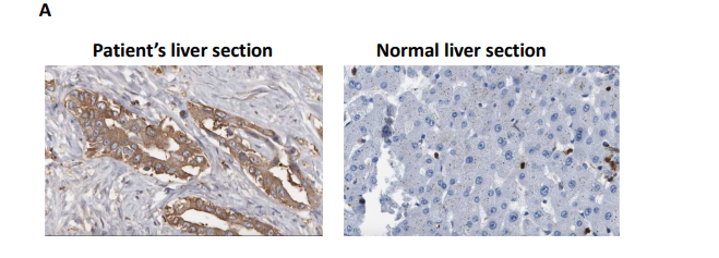
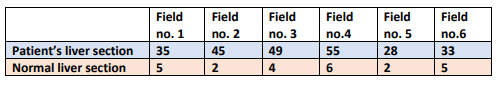
Finally, a liver biopsy taken from the ICC patient at diagnosis was examined by immunohistochemistry using an antibody detecting Ki67 protein. Ki67-positive cells in the patient’s ICC liver tissue were manually counted using microscopy. Results are given in Figure 6.
Buy Answer of This Assessment & Raise Your Grades
Figure 1. Liver enzyme assays, three replicates of each blood sample taken at
different dates.
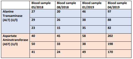
Normal enzyme ranges
Alanine transaminase (U/l): 5–35
Aspartate Aminotransferase (U/l): 7-45
Figure 2: DNA sequence obtained after PCR analysis of DNA extracted from patient’s liver biopsy.
atggccggctacctgcgggtcgtgcgctcgctctgcagagcctcaggctcgcggccggcctgggcgccggcggccctgacagcccccacctcgcaagagcagccgcggcgccactatgccgacaaaaggatcaaggtggcgaagcccgtggtggagatggatggtgatgagatgacccgtattatctggcagttcatcaaggagaagctcatcctgccccacgtggacatccagctaaagtattttgacctcgggctcccaaaccgtgaccagactgatgaccaggtcaccattgactctgcactggccacccagaagtacagtgtggctgtcaagtgtgccaccatcacccctgatgaggcccgtgtggaagagttcaagctgaagaagatgtggaaaagtcccaatggaactatccagaacatcctgggggggactgtcttccgggagcccatcatctgcaaaaacatcccacgcctagtccctggctggaccaagcccatcaccattggcaggcacgcccatggcgaccagtacaaggccacagactttgtggcagaccgggccggcactttcaaaatggtcttcaccccaaaagatggcagtggtgtcaaggagtgggaagtgtacaacttccccgcaggcggcgtgggcatgggcatgtacaacaccgacgagtccatctcaggttttgcgcacagctgcttccagtatgccatccagaagaaatggccgctgtacatgagcaccaagaacaccatactgaaagcctacgatgggcgtttcaaggacatcttccaggagatctttgacaagcactataagaccgacttcgacaagaataagatctggtatgagcaccggctcattgatgacatggtggctcaggtcctcaagtcttcgggtggctttgtgtgggcctgcaagaactatgacggagatgtgcagtcagacatcctggcccagggctttggctcccttggcctgatgacgtccgtcctggtctgccctgatgggaagacgattgaggctgaggccgctcatgggaccgtcacccgccactatcgggagcaccagaagggccggcccaccagcaccaaccccatcgccagcatctttgcctggacacgtggcctggagcaccgggggaagctggatgggaaccaagacctcatcaggtttgcccagatgctggagaaggtgtgcgtggagacggtggagagtggagccatgaccaaggacctggcgggctgcattcacggcctcagcaatgtgaagctgaacgagcacttcctgaacaccacggacttcctcgacaccatcaagagcaacctggacagagccctgggcaggcagtag
Figure 3. IDH2 enzymatic activity detected in untransfected (Parental) cells, cells expressing IDH2 wild-type (WT) or IDH2 Mutant.
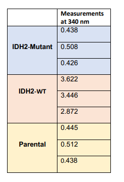
Figure 4. Measurements of NF-kB activity in ICC patient’s liver tissue and normal liver tissue.
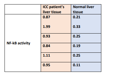
Figure 5. Expression levels of IkBa, B-cell lymphoma 2 (BCL-2), E-Cadherin and vascular endothelial growth factor (VEGF) in IDH2-mutated ICC tumor lysates obtained from patient’s liver biopsy and normal liver.
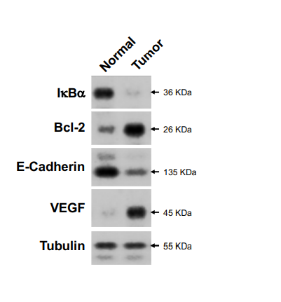
Figure 6. A) Immuno-histochemical staining of Ki67 on normal and patient’s ICC liver tissue. B) Quantification of Ki67 positive cells in the patient sample and normal sample. Six fields were examined for each Ki67 stained slide.
B
Are You Looking for Answer of This Assignment or Essay
Do you need help with your FHEQ level 5 BB2804: Data Analysis Interpretation and Presentation assignment? Our expert team offers excellent online assignment help. Whether you need case study writing services or a written assignment sample for guidance, we’ve got you covered. UK students can pay our professionals for high-quality support tailored to their needs. Get the expert assistance you need to excel in your coursework today!
Answer